
Pathology results are the critical endpoint in preclinical efficacy and safety assessment. The complex structure of eyes makes pathological procedures and examinations a challenge. The difficulty may increase further for those small and diseased animal eyes, and these particular pathology evaluations require unique skills and experience. The TriApex pathology team has skillful technical scientists and experienced diagnostic pathologists, leading the team to conduct ophthalmic pathology assessment and develop innovative methods.
 Introduction of Services
The eyes are generally fixed in Davison fixative and trimmed according to requirement, such as gross lesions or special structures, e.g. the macula, papilla and laser spot. We are able to provide paraffin and frozen slides of mouse, rat, rabbit, Beagle dog and monkey eyes.
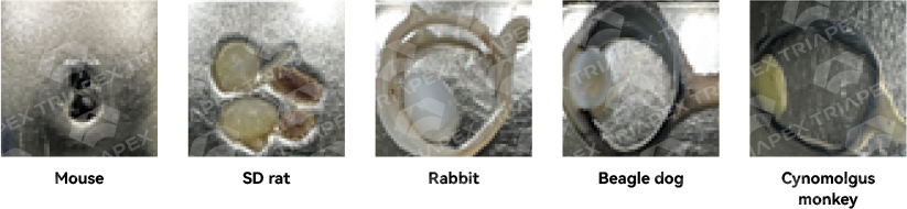
Fig. The eyes were trimmed before embedding in the paraffin.
INHAND diagnostic criteria are applied to diagnose and semi-quantitatively record ocular histological changes. The special disease models some characteristic features of the lesion are recorded.
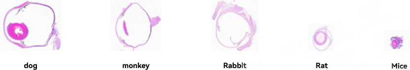
Fig. The eye sections of dog, monkey, rabbit, rat and mouse (from the left to right). Modified from Wang et al (2021). Ophthalmology and therapy, 10(3), 465–494.
Different ocular cells can be specifically stained and analyzed, such as ganglion neurons by flat-mount IHC.
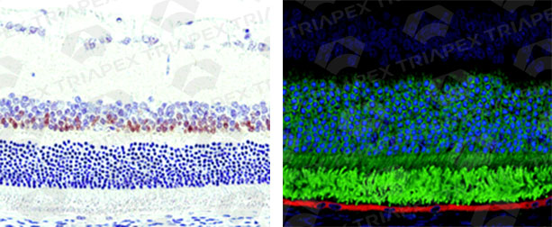
Fig. Specific cells of the eye can be highlighted by using IHC and quantified by using advanced software.
Special stained slides can be scanned in the system and the images are analyzed quantitatively by using simple method or advanced software.
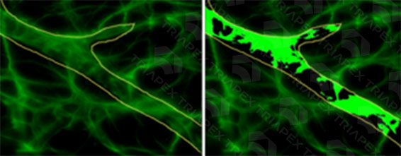
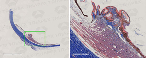
Fig. Quantitative analysis by using advanced software. & Fig. Masson stain and analysis of the ciliary body of cynomolgus monkey


 Official WeChat account
 苏公网安备32011202001081
 Regulatory Policies
 Project Management

 Global


